
For patients who want to self-schedule at their own convenience, click the button above to schedule an appointment now.
Jack W. Bowling Jr., MD
Joint Replacement, Sports Medicine
Board Certified in Orthopedic Surgery, Fellowship Trained in Joint Replacement
Coastal Region
Wilmington – Oleander
Wilmington – Oleander
Coastal Region
Contact
Phone: (910) 332-3800
Physical Therapy Hours
|
Monday: |
8:00AM - 5:00PM |
|
Tuesday: |
8:00AM - 5:00PM |
|
Wednesday: |
8:00AM - 5:00PM |
|
Thursday: |
8:00AM - 5:00PM |
|
Friday: |
8:00AM - 5:00PM |
A native of Gastonia, North Carolina, Jack Bowling, MD, earned a Bachelor of Science in Zoology, graduating summa cum laude from North Carolina State University. He earned his Doctor of Medicine degree from Wake Forest University School of Medicine and stayed to complete his orthopaedic residency at Wake Forest University/Baptist Hospital Medical Center.
Dr. Bowling completed a prestigious fellowship in joint replacement at Central DuPage Hospital/Rush Presbyterian—Saint Luke’s Hospital in Chicago, Illinois. During his fellowship, he mastered a wide range of minimally invasive hip and knee replacement strategies and has continued to lead in various areas of joint replacement, including teaching his peers locally and nationally.
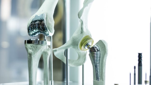
While highly specialized in total joint replacement of the hip, knee and shoulder, Dr. Bowling has also distinguished himself as a leader in arthroscopy of the knee and shoulder. With 20 years of call coverage in Wilmington, he has also gained extensive experience in sports medicine and trauma care, excelling in both acute and reconstructive cases.
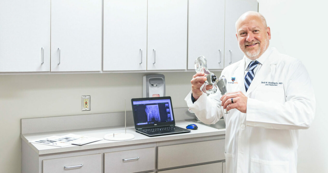
An avid adapter of advanced technology to improve surgical procedures and outcomes, Dr. Bowling uses robotic-assisted technology for joint replacement and is certified using the CORI Robotics surgery system.
Education
- Undergraduate: North Carolina State University
- Medical School: Wake Forest University School of Medicine
- Residency: Wake Forest University/ Baptist Hospital Medical Center
- Fellowship: Central DuPage Hospital/ Rush Presbyterian—Saint Luke’s Hospital
Certifications
- Board-Certified Orthopedic Surgeon
Associations
- American Academy of Orthopedic Surgeons
Affiliations
- Novant Health (NHRMC)
- Wilmington Surgcare
Specialties & Services


Dr. Jack Bowling and Bowling Orthopaedics Join EmergeOrtho Coastal Region's Comprehensive Orthopedic and Spine Practice
Coastal Region
WILMINGTON, NC— On October 1, 2023, EmergeOrtho Coastal Region will welcome Jack W. Bowling Jr., MD, a board-certified, fellowship-trained orthopedic surgeon, and Bowling Orthopaedics to their esteemed team. As founder of Bowling Orthopaedics, Dr. Bowling has been a trusted local provider for 22 years in his Wilmington practice. With extensive experience in minimally invasive joint...
A New Level Of Orthopedic Care Has Emerged
As our patient, you will benefit from a full range of orthopedic services, specialties and technologies, including physical and hand therapy, advanced imaging services, and urgent care walk-in services providing immediate diagnosis and treatment for urgent orthopedic conditions.
Join the EmergeOrtho E-Mail List
Stay informed about the latest orthopedic specialties, news, and upcoming events.


